Mediterranean Diet and Cancer
Epidemiological and other studies have shown that the plant-rich Mediterranean Diet (MD) protects people against cancer and many chronic diseases. MD is characterized by increased consumption of fresh fruits, vegetables, nuts, unrefined grains/legumes, and olive oil, moderate consumption of fish, dairy products, and red wine, and low consumption of red meat.
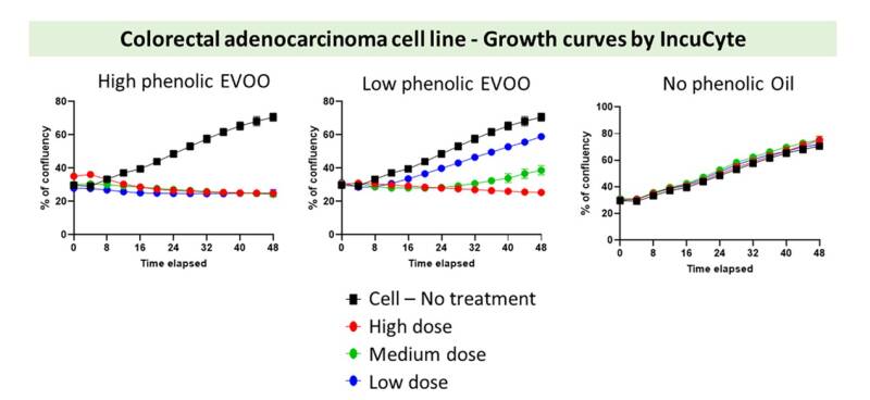
Extra-Virgin Olive Oil (EVOO) contains phenolic compounds that are associated with the beneficials effects of EVOO consumption. Research studies indicate that EVOO rich in phenols has a cardioprotective effect and lower cancer incidence. The aim of our research is to estimate the biological activity of olive oil samples by developing in vitro assays that reconstitute the in vivo activity. The experiments are performed in various cancer cell lines, see figure below.
Wheat is one of the three basic plants (wheat, olive, vine) of the Mediterranean diet and is the best known plant in the history of human nutrition in Europe and the Middle East belonging to the cereals, which are divided into oilseeds, legumes and grains. We examine the effect of increased wheat productivity in comparison to primitive wild native wheat species regarding the anti-cancer effect of their phytochemical/phenolic compounds. The experiments are performed in various cancer cell lines.
Luteolin inhibits tumor growth and angiogenesis
In an attempt to identify phytochemicals contributing to the well-documented preventive effect of plant-based diets on cancer incidence and mortality, we have shown that certain flavonoids inhibit in vitro angiogenesis. We have shown that the flavonoid luteolin inhibited tumor growth and angiogenesis in a murine xenograft model. Furthermore, luteolin inhibited vascular endothelial growth factor (VEGF)-induced in vivo angiogenesis in the rabbit corneal assay. In agreement, luteolin inhibited both VEGF-induced survival and proliferation of human umbilical vein endothelial cells (HUVECs) with an IC(50) of about 5 mumol/L. Luteolin inhibited VEGF-induced phosphatidylinositol 3'-kinase (PI3K) activity in HUVECs, and this inhibition was critical for both the antisurvival and antimitotic affects of the compound. Indeed, luteolin abolished VEGF-induced activation of Akt, a downstream target of PI3K conveying both survival and mitotic downstream signals. Because overexpression of a constitutively active form of Akt rescued HUVECs only from the antisurvival effects of luteolin, the result indicated that luteolin targeted mainly the survival signals of the PI3K/Akt pathway. With regard to its antimitotic activity, luteolin inhibited VEGF-induced phosphorylation of p70 S6 kinase (S6K), a downstream effector of PI3K responsible for G(1) progression.
Eleni Bagli, Maria Stefaniotou, Lucia Morbidelli, Marina Ziche, Konstantinos Psillas, Carol Murphy, Theodore Fotsis; Luteolin Inhibits Vascular Endothelial Growth Factor-Induced Angiogenesis; Inhibition of Endothelial Cell Survival and Proliferation by Targeting Phosphatidylinositol 3′-Kinase Activity. Cancer Res 1 November 2004; 64 (21): 7936–7946. https://doi.org/10.1158/0008-5472.CAN-03-3104

Figure Legend: A Nude mice bearing A-431 tumors (~100 mm³) received daily peritumor injections of luteolin (•, 5 µg/mouse/day) or vehicle (□, 1% EtOH + 1% DMSO) for 7 days. Tumor volume (mm³; mean ± SE, ≥7 mice/group) was determined by caliper measurements. Differences were significant (*P < 0.01, Student’s t test).
B Tumor angiogenesis on day 8 after luteolin (e–h) or vehicle (a–d) treatment. Representative core (a, b, e, f) and periphery (c, d, g, h) sections stained with H&E (a, c, e, g) or anti–ED-B antibody (b, d, f, h). ED-B–positive signals appear in microvessels and remodeling matrix. Magnification ×40.
C Excised tumors were frozen, sectioned (7 µm), and stained with H&E or anti–ED-B antibody. Microvessel density was quantified by counting ED-B–positive vessel-like structures in three sections (seven fields/section, ×20). Data are mean ± SD; *P < 0.05.
The isoflavone metabolite 6-methoxyequol inhibits angiogenesis and suppresses tumor growth
To identify phytochemicals underlying the cancer-preventive effects of plant-based diets, we screened previously untested phytoestrogen metabolites for anti-angiogenic activity, using endothelial cell proliferation as an endpoint. We found that the novel phytoestrogen 6-methoxyequol (6-ME) selectively inhibited VEGF-induced proliferation of human umbilical vein endothelial (HUVE) cells, without affecting their migration or survival. Similarly, 6-ME suppressed FGF-2–induced proliferation of bovine brain capillary endothelial (BBCE) cells. Mechanistically, 6-ME blocked VEGF-induced phosphorylation of ERK1/2 MAPK and its upstream activator MEK1/2, in a dose-dependent manner, while leaving p38 MAPK and AKT phosphorylation unchanged—consistent with its selective effect on proliferation. In vivo, peri-tumor injection of 6-ME in A-431 xenograft tumors significantly reduced tumor growth and neovascularization compared to vehicle-treated controls (P < 0.01).
Bellou S, Karali E, Bagli E, Al-Maharik N, Morbidelli L, Ziche M, Adlercreutz H, Murphy C, Fotsis T. The isoflavone metabolite 6-methoxyequol inhibits angiogenesis and suppresses tumor growth. Mol Cancer. 2012 May 14;11:35. doi: 10.1186/1476-4598-11-35. PMID: 22583931; PMCID: PMC3406996.

Figure Legend: Effect of 6-ME on VEGF-induced endothelial cell survival, migration, tube formation, and phosphorylation of Akt and p38 MAPK.
(A) HUVE cells were serum-starved (5% FBS M199 with heparin and pen/strep) and stimulated with VEGF (50 ng/ml) ± 6-ME (10 μM) for 18 h. Floating and adherent cells were analyzed by flow cytometry; numbers indicate hypodiploid cell percentages (representative of three experiments).
(B) Confluent HUVE monolayers were wounded, serum-starved, and treated with VEGF (50 ng/ml) ± 6-ME (10 μM). Migration was monitored at 37°C, 5% CO₂ using time-lapse microscopy. Graphs show cell number per wound cm ± SD (n = 3).
(C) HUVE cells seeded on Matrigel (8 × 10⁴ cells/ml) were treated with DMSO or 6-ME for 12 h; tube formation was observed by inverted microscopy.
(D) Serum-starved HUVE cells were stimulated with VEGF (50 ng/ml) ± 6-ME for 15 min, lysed, and analyzed by immunoblotting for phospho-Akt, total Akt, phospho-p38, and p38. Data in (A, B) are representative of three independent experiments.
Promotional offer
Don't miss out on the chance to save while enjoying the quality and service you love. Keep an eye on this space for the latest updates and grab these amazing deals while they last!